
Medical imaging represents a transformative field in modern healthcare, empowering clinicians to visualize the internal landscape of the human body without the need for invasive procedures. By converting invisible physiological structures into detailed images, clinicians can diagnose, monitor, and treat a myriad of conditions with precision and confidence. With rapid technological advances, medical imaging continues to evolve, expanding its reach across nearly every medical discipline.
An Overview of Medical Imaging Modalities
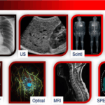
The universe of medical imaging comprises a suite of modalities, each harnessing unique scientific principles to illuminate the complexities of human anatomy and physiology. Below is a summary of the principal modalities, though several other emerging modalities can be expressed later:
| Modality | Primary Principle | Best For |
|---|---|---|
| X-ray Imaging (Radiography, Mammography, etc.) | Ionizing radiation absorption/transmission | Bone injuries, chest and dental imaging |
| Computed Tomography (CT) | Rotating X-ray source & computer reconstruction | Trauma, internal bleeding, cancer detection |
| Magnetic Resonance Imaging (MRI) | Magnetic field and radio frequency waves align hydrogen nuclei | Soft tissue, brain, joints, musculature |
| Ultrasound (US) | High-frequency ultrasound wave reflection | Pregnancy, abdominal organs, blood flow |
| Nuclear Medicine (PET, SPECT) | Radiotracers emitting gamma/positron signals | Cancer, heart disease, functional imaging |
| Electrical Impedance Tomography (EIT) | Electrical current and voltage mapping | Lung, cardiac, and brain monitoring |
| Optical Imaging | Light absorption, scattering, fluorescence | Superficial tissues, functional imaging |
X-ray Imaging (Radiography, Mammography, etc.): X-ray imaging relies on differential absorption of ionizing X-rays as they pass through tissues of varying density. Denser materials like bone absorb more X-rays and appear black, while soft tissues appear white or shades of gray. The method is valued for its speed and clarity, especially in bone and chest investigations. Although it produces 2D images of superimposed organs.
Computed Tomography (CT): Building on traditional X-ray physics, CT scanners capture multiple projections by rotating an X-ray tube around the patient, reconstructing these with advanced software into detailed sequential cross-sectional slices and 3D visuals. This enables clinicians to discern subtle differences in tissue density and create high-resolution images of organs, blood vessels, and bones.
Magnetic Resonance Imaging (MRI): MRI uses powerful magnets to align the hydrogen nuclei in the body, and then perturbs this alignment with radiofrequency pulses with various sequences. As the hydrogen nuclei return to their original state, they emit signals that are processed into exceptional images, notably of soft tissues such as the brain, muscles, heart, and cancers. MRI avoids radiation exposure, making it a preferred modality for numerous conditions.
Ultrasound (US): Ultrasound machines transmit high-frequency acoustic waves via a transducer; as these bounce back from internal structures, they are converted into real-time images. The technique is safe, cost-effective, and portable, widely used for fetal monitoring, abdominal studies, and assessing blood flow with Doppler ultrasound physics.
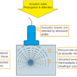
Nuclear Medicine (PET & SPECT): Nuclear medicine visualizes physiological processes by introducing radiotracers, which emit signals captured by specialized cameras. PET scans detect paired gamma photons from positron annihilation, providing metabolic and molecular data vital for cancer staging, while SPECT captures single-photon emissions in various angles around the patient for 3D functional imaging of the heart and brain.
Electrical Impedance Tomography (EIT): EIT, a non-ionizing technology, images the distribution of electrical conductivity within tissues by administering mild electrical currents and measuring resulting voltages. EIT is particularly promising for continuous, bedside lung monitoring and is evolving for real-time assessment of cardiac and neurological function.
Optical Imaging: Using light-based techniques such as endoscopy, optical coherence tomography, and fluorescence imaging, this modality is non-invasive and excels at visualizing superficial organs, vasculature, and molecular processes at high spatial resolutions.
Leading Medical Imaging Equipment Manufacturers and Their Devices
Global medical imaging is propelled by renowned manufacturers that continually innovate to improve image quality, reliability, and patient safety. The dominant industry players, recognized for their extensive expertise and global reach, include:
- Siemens Healthineers: Offers an advanced range from digital X-ray, CT, and MRI scanners to PET/CT hybrid systems and high-end ultrasound machines. Siemens prioritizes automation and artificial intelligence integration for workflow optimization.
- GE Healthcare: Designs a broad spectrum of devices across all modalities, including compact ultrasound for point-of-care and powerful MRI and CT systems known for their speed and image clarity. Their nuclear medicine platforms lead in molecular imaging.
- Philips Healthcare: Known for ergonomic, patient-centered solutions, Philips manufactures MRI, ultrasound, and digital X-ray systems as well as digital mammography and hybrid imaging units.
- Canon Medical Systems (formerly Toshiba): Focused on user-friendly CT, ultrasound, and MRI devices, renowned for durability and advanced image reconstruction capabilities.
- Hitachi Healthcare: Distinguished by innovative MRI systems with strong patient comfort features, digital ultrasound devices, and high-quality CT systems.
- Fujifilm: A frontrunner in digital radiography, mammography, and ultrasound, offering compact, portable devices for versatile clinical applications.
These manufacturers have tailored their products to the evolving needs of hospitals and clinics, supporting the movement toward digital and connected healthcare with robust IT, cybersecurity, and training support. Their work ensures that imaging hardware remains accessible and reliable for both routine diagnostics and high-acuity care.
Specialties and Professionals in the Medical Imaging Sector
Medical imaging is a collaborative ecosystem, combining the expertise of specialized professionals to ensure high-quality patient care. The principal roles include:
- Radiologists: These physicians lead in interpreting imaging studies, recommending further tests, and collaborating in treatment planning. Their diagnostic acumen is key to accurate patient management.
- Radiographers / Radiologic Technologists: Highly trained in operating imaging equipment and optimizing imaging protocols for quality and safety, these allied professionals acquire images and ensure patient well-being throughout the imaging procedure.
- Medical Physicists: Experts in the application of physics to imaging, physicists maintain equipment quality and safety, participate in device calibration, and innovate in radiation protection and image optimization. Medical physicists bridge the gap between technological innovation and clinical application.
- Medical Imaging Assistants: Support imaging professionals with patient preparation, data management, and workflow facilitation, especially in large departments.
- Biomedical Engineers: With close collaboration with medical physicists, design and maintain electrical and mechanical parts of sophisticated imaging technology.
- Nuclear Medicine Technologists: Specialize in handling radiopharmaceuticals, performing PET/SPECT imaging, and ensuring regulatory compliance in radiation safety.
- Cardiac and Vascular Sonographers: Experts in using ultrasound to assess heart and blood vessel function, vital in cardiology and vascular surgery.
- Researchers and Data Scientists: Involved in advancing imaging technology, artificial intelligence, image processing, and applications of big data and machine learning to medical imaging.
This multidisciplinary framework supports the safe and efficient delivery of imaging services, underpinned by continuous professional development due to rapid technological change.
The Physics and Principles Behind Each Modality
A deep understanding of physics is fundamental to every aspect of medical imaging. Each modality leverages a unique scientific phenomenon to capture visual data:
- X-ray Imaging: X-rays – generated when high-energy electrons strike a high atomic number metal target, Tungsten, Molybdenum, Rhodium, etc.- pass through the patient and are differentially absorbed by dense tissues. The remaining photons are detected and converted into an image, with bones appearing darker due to higher absorption.
- CT: Like X-ray, CT uses ionizing radiation but rotates the X-ray source around the patient, collecting a multitude of projection images that are mathematically reconstructed into cross-sectional ‘slices’ using algorithms such as filtered back-projection or iterative reconstruction. For more information about detection mechanisms and CT reconstruction techniques, my posts here and here can be read.
- MRI: MRI operates on the principle of nuclear magnetic resonance, where hydrogen nuclei align in an external magnetic field. Radiofrequency pulses momentarily disturb this alignment, and as nuclei return to their original orientation, they emit signals that are mapped into images. Pulse sequences and magnetic field gradients allow for 3D image reconstruction and extraordinary flexibility in image contrast and spatial resolution.
- Ultrasound Imaging: Ultrasound uses piezoelectric crystals to create ultrasound waves that travel through tissue and reflect off internal structures. The returning echoes are translated into dynamic images, with Doppler technology enabling assessment of blood flow by measuring frequency shifts caused by movement.
- PET and SPECT: These modalities use injected radiotracers that emit gamma rays (SPECT: single photons, PET: pairs of photons in opposite directions following positron-electron annihilation). Ring-shaped or rotating detectors capture these emissions, and advanced software reconstructs three-dimensional maps of tracer distribution, reflecting metabolic or functional activity.
- EIT: Small alternating currents are applied through surface electrodes, and voltage differences are measured around the body. By solving complex inverse problems, images reflecting tissue conductivity are generated, providing insight into functional processes like ventilation distribution.
- Optical Imaging: Light, often in the visible or infrared spectrum, passes into tissue where it is scattered and absorbed. Detectors measure transmitted or reflected light, exploiting differences in optical properties to image structure or function, particularly in superficial organs.
Unique Advantages and Limitations of Each Modality
| Modality | Strengths | Limitations |
|---|---|---|
| X-ray | Fast, simple, low cost | Ionizing radiation, limited soft tissue contrast |
| CT | Rapid, high-resolution, 3D images | Radiation dose; limited soft tissue contrast |
| MRI | No ionizing radiation, high soft tissue contrast | Expensive; time-consuming; contraindicated with some implants |
| Ultrasound | Real time, noninvasive, portable, safe | Limited by bone/air interfaces; operator-dependent |
| PET/SPECT | Quantitative functional imaging | Radiation, expensive, limited spatial resolution |
| EIT | Safe, bedside, continuous monitoring | Lower spatial resolution, still under development |
| Optical Imaging | High resolution for superficial structures | Limited penetration depth |
Final Thoughts
Medical imaging is a vital pillar of contemporary healthcare, integrating advanced devices, specialized expertise, and profound physical principles to deliver life-saving diagnostics and guide treatment. From the rapid assessment of trauma with CT to the monitoring of fetal development via ultrasound, the careful orchestration of technology and skill improves patient outcomes worldwide. As innovation marches forward—driven by leading manufacturers, dedicated professionals, and a culture of safety and research—medical imaging will only expand its indispensable role in medicine.


Leave a Reply