
Photoacoustic Imaging (PAI) is a hybrid imaging technology that usually utilizes a pulsed optical source and an ultrasound probe for signal generation and detection. It is known for its capability in the acquisition of optical contrast and penetration depths much higher than pure optical imaging modalities, due to the use of ultrasound wave propagation.
The Photoacoustic (PA) effect
Basically, the PA effect refers to the phenomenon in which an optical wave illuminates a target, and consequently, acoustic waves are generated. This phenomenon was first discovered by Alexander Graham Bell during his photophone experiments, which transmitted speech to a remote listener. The photophone used vibrating mirrors to modulate a sunlight beam with voice signals, then transformed the signals back into sound at the receiving mirror. Following this invention, he stated that illuminating a solid material via sunlight produced acoustic signals at a modulated frequency. However, this phenomenon did not come into account till the discovery of the laser.
The PA effect primarily operates based on the absorption of light and the subsequent pressure changes in the material. This involves several stages, including light absorption, thermal expansion, and acoustic wave generation. The first step involves the absorption of light by a material. The light absorbance of materials depends on their absorption coefficients. The absorbed light energy is transformed into heat, increasing the local temperature. As the temperature rises, the material expands. This rapid thermal expansion generates localized pressure changes in the medium surrounding the absorbing material. The expansion occurs because the absorbed energy causes the molecules within the material to vibrate more energetically. The pressure changes caused by thermal expansion create acoustic waves. These waves propagate through the material and can be detected by appropriate sensors. The characteristics of these acoustic waves depend on several parameters, including the intensity of the light and the material properties.
Signal generation and detection in Photoacoustic imaging
For signal generation in PAI, the pulsed optical source with a suitable pulse duration illuminates the tissue of interest. The laser pulse duration must be shorter than the tissue or medium’s thermal and stress relaxation times. This condition ensures that the absorbed energy does not disperse away before it can generate a pressure wave. Thermal relaxation time is the time it takes for the absorbed heat to diffuse through the medium, and stress relaxation time is the time it takes for the mechanical stress caused by the absorption of the laser energy to relax.
Since biological tissues have higher absorption coefficients in the near-infrared or visible part of the spectrum, and light penetrates more in these windows, PAI primarily works well in this part of the spectrum. Since each tissue has a unique optical absorption at a specified wavelength, the contrast in PA images can be defined.
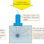
When a pulsed laser irradiates the tissue, photons scatter and are absorbed in chromophores (As shown in the Figure). The deposited energy should be confined locally and be able to generate a strong pressure wave. Absorbed energy at the location r of a chromophore, H(r) depends on μ_a and illuminated flux at the location r (Equation 1);
H(r)=μ_a (r)F(r,μ_a,μ_s,g) Equation 1
Where F(r, μa, μs, g) is the light fluence.
Under the mentioned circumstances, the absorbed energy at location r leads to the generation of an initial pressure wave P_0, which then propagates through the tissue. These acoustic waves are eventually detected by a sensor, which can be a typical ultrasound transducer. The magnitude of the generated initial PA pressure can be described as follows:
P_0=(β〖v_s〗^2)/c_p H(r) Equation 2
Where β is the thermoelastic coefficient of volume expansion, and c_p is the specific heat capacity. Then, acoustic waves travel through the medium and are detected by a transducer.
Briefly, in a PA imaging system, the pulsed laser with an appropriate wavelength corresponding to the peak absorption coefficient of the target tissue illuminates the tissue. The light absorption leads to subsequent heating, which, due to the thermoelastic effect, causes ultrasound waves to be produced and propagate through the medium to reach a transducer; these processes have been shown in the Figure at the top of this post.
A common excitation source for PAI is solid-state lasers, such as Q-switched rare-earth-doped crystals (e.g., Nd: YAG). These sources provide high pulse energy, resulting in higher Signal-to-Noise Ratios (SNRs). Despite their perfect ability to provide high-quality images, they tend to be expensive, bulky, and require cooling, making them less suitable for clinical use. Consequently, semiconductor-based sources are increasingly being adopted. Semiconductor sources, including diode lasers and LEDs, can be used in surgical units or at the patient’s bedside as they are cheap, lightweight, and portable. Although these sources provide flexibility in the output wave shape, their energy is less than Nd: YAG lasers. Fiber lasers can be an alternative to diode lasers and LEDs. They operate based on the amplification method, are more compact, and provide higher output; however, additional research is needed to utilize them.
Generated PA waves are mostly detected by a typical ultrasound probe, which translates the pressure of detected waves to an electrical current based on the piezoelectric effect. The conventional probes are convenient and highly sensitive; however, new probes based on capacitive micromachined ultrasound transducers can also work in wider bandwidths, which causes the imaging system to be more sensitive and provides higher resolution. Moreover, other types of transducers, such as ultra-wideband or focused transducers, have also recently drawn attention. Also, geometry and the number of transducer elements are two important factors in PA signal detection. The higher number of elements results in higher resolution. Possible geometries include planar, spherical, concave, arc, and ring-shaped designs. Although planar transducers are the most widely used, they can cause some artifacts. Moreover, detectors with unique shapes exist for specific applications; for example, the developed hockey stick-shaped sensor for dental imaging.
If you need more technical information about fundamentals and applications of Photoacoustic Imaging, I suggest reading these sources:
Fundamentals of Photoacoustic Imaging, published by Springer
Applications of Photoacoustic Imaging in Preclinical Research, published by Springer
I hope this post can help you in this amazing field. For further discussion, you can contact me.


Leave a Reply