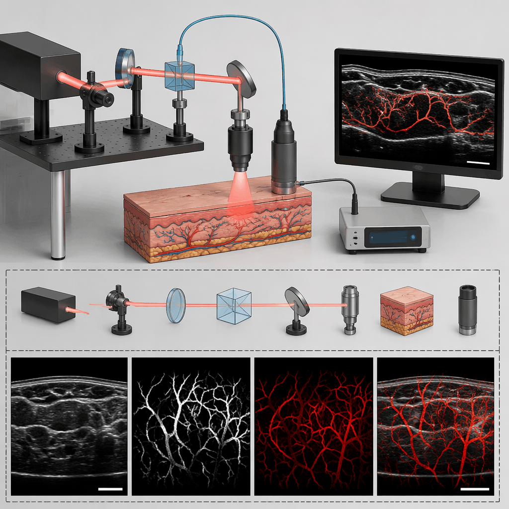
If you have been following advances in label-free optical microscopy, you may have come across two terms that seem confusingly similar: photoacoustic remote sensing (PARS) and photon absorption remote sensing (also PARS). Both come from the same lab (PhotoMedicine Labs), both share the same acronym, and both address non-contact absorption imaging. So are they the same concept? The short answer is: they are not two separate inventions, but rather a single technology at different stages of evolution. Understanding how one grew out of the other reveals a story of scientific ambition, from a clever optical trick for detecting photoacoustic pressure to a comprehensive microscope that watches the entire photon absorption event unfold.
The First Generation: Photoacoustic Remote Sensing
In 2017, Parsin Haji Reza and colleagues published a landmark paper in Light: Science & Applications introducing photoacoustic remote sensing (PARS) microscopy. The core idea was deceptively simple yet powerful: instead of placing a piezoelectric ultrasound transducer in contact with the sample (as conventional photoacoustic microscopy requires), why not detect the initial photoacoustic pressure optically, right where it is generated?
In PARS, a pulsed excitation laser is focused into a sample, and a co-focused continuous-wave probe beam monitors the elasto-optic refractive index modulations produced by thermo-elastic pressure transients at the subsurface origin where pressures are largest. Because the probe beam relies on intensity-reflectivity changes rather than interferometric phase measurement, the architecture is non-interferometric, precluding detection of surface oscillations and other phase-modulation phenomena. The result is a non-contact, reflection-mode photoacoustic microscope with cellular-scale resolution and centimeter-scale working distance.
This first-generation PARS excelled at what it was designed for: capturing non-radiative relaxation, the energy a molecule sheds as heat and pressure after absorbing a photon. Over the following years, the technology was extended with lock-in amplification for improved sensitivity, coherence-gated depth resolution, real-time functional imaging with multi-wavelength excitation via stimulated Raman scattering in optical fiber, hyperspectral absorption spectroscopy from 210 nm to 1550 nm, and chromophore-selective imaging of unstained human tissues.
The Missing Half of the Story
Despite its success, first-generation PARS shared a fundamental limitation with all purely photoacoustic or purely photothermal modalities: it captured only the non-radiative fraction of each absorption event. When a molecule absorbs a photon, it can de-excite along two dominant pathways: (1) non-radiative relaxation, shedding energy as heat and pressure, and (2) radiative relaxation, re-emitting energy as fluorescence or other optical emissions. The relative contribution of each path depends on the molecule’s quantum yield and local environment. Hemoglobin and nucleic acids, for instance, have near-zero quantum yield, meaning almost all their absorbed energy is released non-radiatively. Connective-tissue proteins like collagen and elastin, in contrast, exhibit appreciable autofluorescence.
A purely non-radiative modality simply cannot see biomolecules that relax predominantly through radiative channels, and vice versa. This creates blind spots that limit specificity, a serious drawback for applications such as label-free histopathology, where nuclear and stromal structures must be visualized simultaneously.
The Second Generation: Total-Absorption PARS
To overcome this limitation, the group introduced what they called total-absorption photoacoustic remote sensing (TA-PARS) around 2021-2022. TA-PARS added a dedicated radiative detection pathway alongside the existing non-radiative probe. After each excitation pulse, Stokes-shifted fluorescent photons emitted by the sample are spectrally filtered and directed to an avalanche photodiode, while the confocal probe beam simultaneously records photothermal and photoacoustic-induced intensity modulations.
With both pathways captured from the same excitation event, TA-PARS introduced two novel contrast metrics: Total Absorption (TA), the sum of the radiative and non-radiative signals, which reveals a molecule’s true optical absorption independent of mechanism-specific efficiency factors; and the Quantum Efficiency Ratio (QER), the differential ratio of radiative to non-radiative relaxation, which encodes chromophore-specific attributes correlated with, but not equivalent to, the fluorescence quantum yield. Empirical validation across fluorescent dyes showed a strong correlation (R = 0.988) between the QER and known quantum efficiencies.
TA-PARS demonstrated label-free H&E-like histological images in human skin, breast, and brain tissues, with sufficient contrast to identify individual atypical nuclei and connective tissue structures from a single acquisition.
From “Photoacoustic” to “Photon Absorption”: A Name That Matches the Science
By 2023-2024, it became clear that the term “photoacoustic remote sensing” no longer accurately described the technology. The modality was no longer sensing only photoacoustic (pressure) signals; it was capturing the entire photon-absorption interaction, including radiative emissions, photothermal transients, photoacoustic pressures, optical scattering, and attenuation. The name “photoacoustic” had become an underspecification.
In their 2024 comprehensive review, the group formally introduced the name Photon Absorption Remote Sensing (PARS), explicitly stating: “In this manuscript, we introduce Photon Absorption Remote Sensing (PARS), a comprehensive optical absorption microscopy technique which has evolved from a photoacoustic remote sensing method previously proposed by our group”. Similarly, the 2023 vascular imaging paper notes: “PARS has evolved from photoacoustic remote sensing reported by Haji Reza et al.”.
The PARS acronym was deliberately preserved. But the full name now reflects what the technology actually does: it remotely senses all dominant photon absorption processes at each excitation event. The earlier name described a subset of the capability; the new name describes the whole.
How the Mechanism Expanded
To appreciate the scale of the conceptual expansion, consider what a PARS acquisition now records at each pixel :
- Excitation: A pulsed laser delivers photons to the specimen.
- Non-radiative detection: A confocal probe beam captures transient temperature (photothermal) and pressure (photoacoustic) induced optical modulations, providing contrast from molecules like hemoglobin and DNA that have low quantum yield.
- Radiative detection: Emitted Stokes-shifted photons are collected and measured, providing contrast from fluorophores like collagen, elastin, and NADH.
- Scattering and attenuation: The unmodulated reflections or transmissions of the excitation and detection beams provide structural and morphological context.
From these four channels, PARS computes the Total Absorption (TA) and Quantum Efficiency Ratio (QER), offering biomolecule-specific contrast that neither radiative nor non-radiative modalities can achieve independently. Any molecule that absorbs light will contribute some signal to PARS, either through its radiative or non-radiative channel, effectively eliminating the quantum-yield blind spot that constrains fluorescence-only or photoacoustic-only techniques.
What Changed and What Stayed the Same
The table below summarizes the evolution across the three conceptual stages:
| Feature | Photoacoustic RS (1st Gen) | TA-PARS (2nd Gen) | Photon Absorption RS (Current) |
|---|---|---|---|
| Non-radiative contrast | Yes | Yes | Yes |
| Radiative contrast | No | Yes | Yes |
| Scattering/attenuation | Partial | Yes | Yes |
| Total Absorption (TA) | No | Yes | Yes |
| Quantum Efficiency Ratio (QER) | No | Yes | Yes |
| Non-contact operation | Yes | Yes | Yes |
| Working distance | >2 cm | >1 cm | >1 cm |
| Label-free | Yes | Yes | Yes |
| Core architecture | Pump-probe | Pump-probe + fluorescence pathway | Pump-probe + fluorescence pathway + spectroscopy |
The foundational pump-probe architecture, the all-optical non-contact operation, and the core principle of detecting photoacoustic-induced refractive-index changes at the excitation origin remain unchanged. What changed is the addition of a radiative detection channel and, with it, a fundamental broadening of what “PARS” means.
The Patent Trail: Protecting a Technology Through Its Evolution
The patent portfolio reflects the same arc of expanding scope. The earliest filings, dating to a 2014 priority date, protect the foundational concept of optically detecting photoacoustic signals without acoustic coupling. Later filings protect extensions such as camera-based detection, single-source configurations, and eventually the full photon-absorption system with radiative detection and machine-learning processing.
For researchers and clinicians evaluating PARS technology, the key takeaway is this: if you encounter the term “photoacoustic remote sensing” in older literature, it refers to the non-radiative-only first generation. If you see “photon absorption remote sensing” in newer work, it refers to the comprehensive, current-generation technology that captures the full absorption interaction. Both share the same acronym, the same core detection mechanism for non-radiative signals, and the same non-contact, label-free philosophy. The newer name is not a rebranding for marketing purposes; it is a scientific re-specification that matches a genuinely expanded capability.
At Professor Haji Reza’s PhotoMedicine Labs at the University of Waterloo and through the spinoff company IllumiSonics, the technology continues to advance toward clinical translation, with recent work demonstrating diagnostic equivalence to gold-standard H&E staining in skin cancer biopsies and automated whole-slide imaging pipelines.
References
Patents
- US10,117,583 B2 – P. Haji Reza, R. Zemp, “Photoacoustic remote sensing (PARS),” priority date 2014-10-22, granted 2018-11-06.
- US10,682,061 B2 – IllumiSonics Inc., “Photoacoustic remote sensing (PARS)” (continuation), filed 2018-08-29, granted 2020-06-16.
- US11,298,027 B2 – IllumiSonics Inc., “Photoacoustic remote sensing (PARS)” (continuation), filed 2020-04-13, granted 2022-04-12.
- US12,207,902 B2 – IllumiSonics Inc., “Photoacoustic remote sensing (PARS)” (continuation), filed 2022-03-14, granted 2025-01-28.
- US10,327,646 B2 – P. Haji Reza, “Non-interferometric photoacoustic remote sensing (NI-PARS),” filed 2016-02-02, granted 2019-06-25.
- US11,517,202 B2 – IllumiSonics Inc., “Non-interferometric photoacoustic remote sensing (NI-PARS),” filed 2016-02-02, granted 2022-12-06.
- US10,627,338 B2 / US11,022,540 B2 – IllumiSonics Inc., “Camera-based photoacoustic remote sensing (C-PARS),” filed 2017-03-23.
- US11,950,882 B2 – K. Bell, P. Haji Reza, “Single source photoacoustic remote sensing (SS-PARS),” filed 2019-03-15, granted 2023-01-31.
- US11,841,315 B2 – IllumiSonics Inc., “Photoacoustic remote sensing (PARS), and related methods of use,” filed 2019-12-19, granted 2023-12-12.
- US11,122,978 B1 – IllumiSonics Inc., “PARS imaging methods,” filed 2020-06-18, granted 2021-09-21.
- US11,786,128 B2 – IllumiSonics Inc., “PARS imaging methods,” filed 2020-06-18, granted 2023-10-17.
- WO2021255695A1 / EP4337934A4 – IllumiSonics Inc., P. Haji Reza, K. Bell, B. Ecclestone et al., “Photoabsorption remote sensing imaging,” priority date 2021-05-12.
- US12,100,153 B2 – B. Ecclestone, J. Tweel, M. Bishop, P. Haji Reza / IllumiSonics Inc., “Photon absorption remote sensing system for histological assessment of tissues,” filed 2024-02-08, granted 2024-09-24.
- CA3272908A1 – P. Haji Reza, B. R. Ecclestone, J. E. D. Tweel et al. / IllumiSonics Inc., “Machine-learning processing for photon absorption remote sensing signals,” filed 2023-11-09.
- CA3161943A1 – P. Haji Reza, K. Bell, “Photoacoustic remote sensing (PARS), and related methods,” filed 2017.
Selected Publications
- P. Haji Reza, W. Shi, K. Bell, R. J. Paproski, R. J. Zemp, “Non-interferometric photoacoustic remote sensing microscopy,” Light: Science & Applications, 6(1), e16278, 2017.
- K. Bell, P. Haji Reza, W. Shi, R. J. Zemp, “Temporal evolution of low-coherence reflectometry signals in photoacoustic remote sensing microscopy,” Applied Optics, 56(18), 5172-5178, 2017.
- K. Bell, P. Haji Reza, R. J. Zemp, “Coherence-gated photoacoustic remote sensing microscopy,” Optics Express, 26(18), 23689-23701, 2018.
- K. Bell, P. Haji Reza, R. J. Zemp, “Real-time functional photoacoustic remote sensing microscopy,” Optics Letters, 44(14), 3466-3469, 2019.
- S. Abbasi, M. Le, B. Sonier, K. Bell, D. Dinakaran et al., “Chromophore selective multi-wavelength photoacoustic remote sensing of unstained human tissues,” Biomedical Optics Express, 10(11), 5461-5470, 2019.
- K. Bell, P. Haji Reza, “Non-contact reflection-mode optical absorption spectroscopy using photoacoustic remote sensing,” Optics Letters, 45(13), 3427-3430, 2020.
- P. Kedarisetti, N. J. M. Haven, B. S. Restall, M. T. Martell, R. J. Zemp, “Label-free lipid contrast imaging using non-contact near-infrared photoacoustic remote sensing microscopy,” Optics Letters, 45(16), 4559-4562, 2020.
- M. T. Martell, N. J. M. Haven, R. J. Zemp, “Multimodal imaging with spectral-domain optical coherence tomography and photoacoustic remote sensing microscopy,” Optics Letters, 45(17), 4859-4862, 2020.
- K. Bell, L. Mukhangaliyeva, L. Khalili, P. Haji Reza, “Hyperspectral absorption microscopy using photoacoustic remote sensing,” Optics Express, 29(15), 24338-24352, 2021.
- N. Pellegrino, B. R. Ecclestone, P. Fieguth, P. Haji Reza, “Time-domain feature extraction for target specificity in photoacoustic remote sensing microscopy,” Optics Letters, 47(15), 3952-3955, 2022.
- B. R. Ecclestone, K. Bell, S. Sparkes, D. Dinakaran, J. R. Mackey, P. Haji Reza, “Label-free complete absorption microscopy using second-generation photoacoustic remote sensing,” Scientific Reports, 12, 16829, 2022.
- J. E. D. Tweel et al., “Photon Absorption Remote Sensing Imaging of Breast Needle Core Biopsies,” Journal of Clinical Medicine, 30(11), 708, 2023.
- J. A. Simmons, S. J. Werezak, B. R. Ecclestone, J. E. D. Tweel, H. Gaouda et al., “Label-Free Non-Contact Structural and Functional Vascular Imaging using Photon Absorption Remote Sensing,” arXiv:2310.05260, 2023.
- B. R. Ecclestone, A. Tummon Simmons, J. Tweel, C. Kaur, A. Hajiahmadi, P. Haji Reza, “Photon Absorption Remote Sensing (PARS): A Comprehensive Approach to Label-free Absorption Microscopy Across Biological Scales,” arXiv:2403.04229 / Journal of the Optical Society of America B, 43(3), A74, 2026.
- B. R. Ecclestone, J. A. Simmons, J. E. D. Tweel, D. Dinakaran, P. Haji Reza, “Photon Absorption Remote Sensing (PARS): Comprehensive Absorption Imaging Enabling Label-Free Biomolecule Characterization and Mapping,” Scientific Reports, 2026.
- B. R. Ecclestone, J. E. D. Tweel, M. Abi Daoud, H. Gaouda, D. Dinakaran, P. Haji Reza, “Photon Absorption Remote Sensing Virtual Histopathology: A Preliminary Exploration of Diagnostic Equivalence to Gold-Standard H&E Staining in Skin Cancer Excisional Biopsies,” arXiv:2504.18737, 2025.
- J. Dhillon, C. Kaur, J. E. D. Tweel, B. Ecclestone, J. Tummon Simmons, P. Haji Reza, “Toward an optical spectroscopy technique using photon absorption remote sensing,” Journal of the Optical Society of America B, 43(3), A206, 2026.
- C. Kaur, A. Hajiahmadi, B. Ecclestone, J. Tweel, J. E. Simmons, P. Haji Reza, “Investigation of Non-Radiative Relaxation Dynamics Under Pulsed Excitation Using Photon Absorption Remote Sensing: A Proof-of-Principle Study in Mechanical Sensing,” arXiv:2502.19650, 2025.
- K. Bell, “Photoacoustic Remote Sensing (PARS) Microscopy,” PhD Dissertation, University of Alberta, 2019.
- N. Pellegrino, “Developments Towards Chromophore-Selectivity in Photoacoustic Remote Sensing Microscopy,” MSc Thesis, University of Waterloo, 2022.
- B. Ecclestone, “Label-free optical microscopy: Photon Absorption Remote Sensing (PARS),” PhD Dissertation, University of Waterloo, 2026.


Leave a Reply